|
|
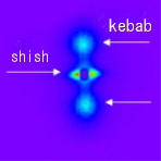
Nano-structure
ーshishkebab structureー
|
 |
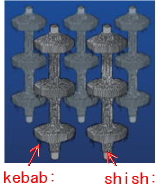
Lamella of folded chain crystal
|
|
|
Polyethylene fiber
(1mmφ240kg)
|
|
|
|
Neutron scattering with deuterium labeling
and X-ray scattering
Role of high molecular weight component
|
 |
 |
|
neutron
|
X-ray
|
|
|
|

|
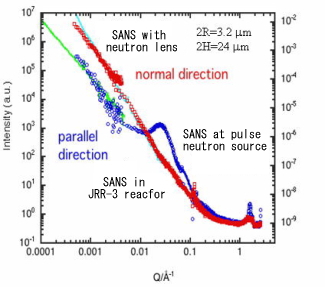
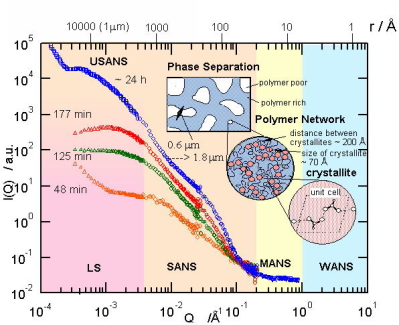
Hierarchic structure by neutron scattering
|
Measurements with SANS with neutron lens、pulse neutron SANS、reactor neutron
SANS
|
|
|
I(Q) over 4 decades (left) and Schematic sketch of Hierarchic structure
of shish-kebab (right)
|
Polymer, 46, 1878 (2005)
Polymer, 47, 5669 (2006)
Macromolecules, 39, 7617 (2006)
Macromolecules, submitted
|
|
|
|
|
|
Structure Change Correlated with Dynamics
in Glassy State
|
|
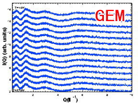
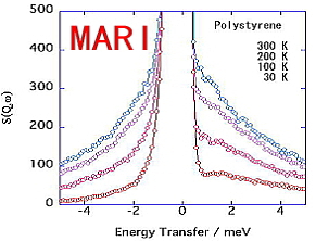
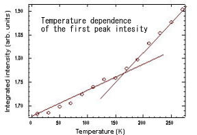
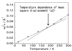
We have studied structure change in glassy deuterated polystyrene
with temperature, and found that the temperature dependence of the first
peak intensity changes at around 170 K, corresponding to the dynamical
change at around 180 K at which the so-called picosecond fast process appears.
|
Structure factor S(Q) of deuterated
polystyrene measured by GEM
|
Dynamic scattering las S(Q,w)
of polystyrene measured
by MARI
|
 |
 |
 |
 |
| Structure change corresponding to the dynamic change at =170K
|
|
Onset of fast process at around 180 K
|
|
|
Hierarchic Structure of Poly(vinyl alcohol) Gel
|
We have studied hierarchic structure of water-soluble and biocompatible
PVA gels using wide-angel, small-angle and ultra-small-angle neutron scattering
and light scattering in a wide Q range from 10^-4 to 10 A^-1. It
revealed structure due to phase separation in micrometer order, network
structure of nm scale and structure of cross-linking points in A order.
|
 |
 |
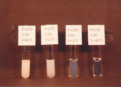
| Appearance of PVA gels prepared in DMSO/water (60/4) at various temperatures.
|
|
|
|
|
|
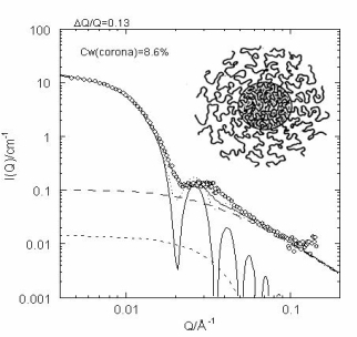
Structure of Polymer Micelles
by SANS with Deuterium Labeling Method
|
When diblock copolymers are added in a selective solvent, aggregation occurs
to form polymer micelles in analogous to micellization of surfactant molecules.
Such polymeric micelles are isolated in a dilute region while they form
a lattice in a concentrated region. In the dilute solution, the polymer
chains in the corona part of the micelle can be regarded as tethered chains
on a surface of sphere. In this work we have investigated the structure
of polymer micelle of polybutadiene-deuterated polystyrene diblock copolymer
in deuterated n-decane (deuterated labeling method), so that we have
the corona contrast. The data were analyzed in term of hard core-shell
model, but blob scattering form polymer chains in the corona was necessary
to describe the whole scattering profile.
|
|
Corona contrast
 |
Analysis in terms
of core-shell model
|
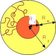 |
Agg. No = 78.7
R2 = 219 A
R1 = 68 A
RB = 151 A
|
|
| I(Q) from polybutadiene-deterated polystyrene diblock polymer micelle and the results of analysis.
|
|
J. Chem. Phys., 122, 144905 (2005)
|
|
|